What is Stress Echocardiography?
A stress echocardiography, also called an echocardiography stress test or stress echo, is a procedure that determines how well your heart and blood vessels are working.
During a stress echocardiography, you’ll exercise on a treadmill or stationary bike while your doctor monitors your blood pressure and heart rhythm.
When your heart rate reaches peak levels, your doctor will take ultrasound images of your heart to determine whether your heart muscles are getting enough blood and oxygen while you exercise.
Your doctor may order a stress echocardiography test if you have chest pain that they think is due to coronary artery disease or a myocardial infarction, which is a heart attack. This test also determines how much exercise you can safely tolerate if you’re in cardiac rehabilitation.
The test can also tell your doctor how well treatments such as bypass grafting, angioplasty, and anti-anginal or antiarrhythmic medications are working.
This test is safe and noninvasive. Complications are rare, but can include:
- An abnormal heart rhythm.
- Dizziness or Fainting.
- Heart attack.
This test usually occurs in an echocardiography laboratory, or echo lab, but it can also occur in your doctor’s office or other medical setting. It normally takes between 45 and 60 minutes.
Before you take the test, you should do the following:
-
- Make sure not to eat or drink anything for three to four hours before the test.
- Don’t smoke on the day of the test because nicotine can interfere with your heart rate.
- Don’t drink coffee or take any medications that contain caffeine without checking with your doctor.
- If you take medications, ask your doctor whether you should take them on the day of the test. You shouldn’t take certain heart medications, such as beta-blockers, isosorbide-dinitrate, isosorbide-mononitrate (Isordil Titradose), and nitroglycerin, before the test. Let your doctor know if you take medication to control diabetes as well.
- Wear comfortable, loose-fitting clothes. Because you will exercise, make sure to wear good walking or running shoes.
Resting Echocardiography.
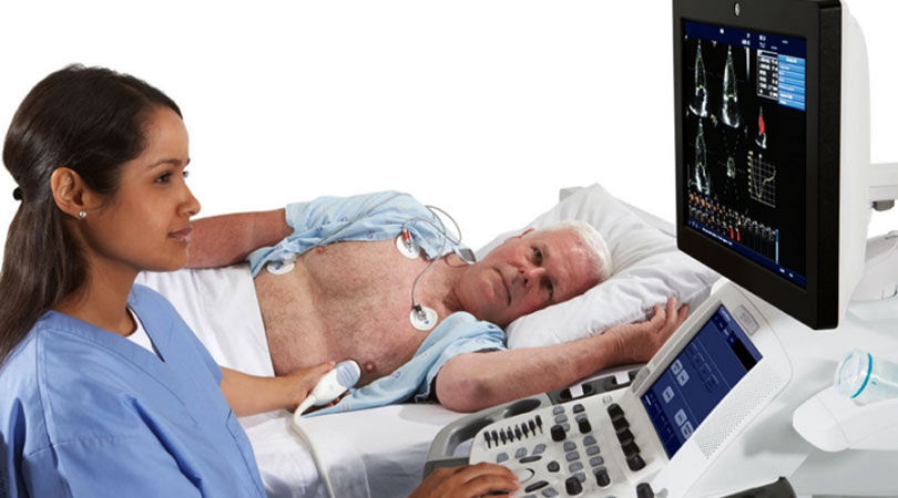
Your doctor needs to see how your heart functions while you’re at rest to get an accurate idea of how it’s working. Your doctor begins by placing 10 small, sticky patches called electrodes on your chest. The electrodes connect to an electrocardiograph (ECG).
The ECG measures your heart’s electrical activity, especially the rate and regularity of your heartbeats. You’ll likely have your blood pressure taken throughout the test as well.
Next, you’ll lie on your side, and your doctor will do a resting echocardiogram, or ultrasound, of your heart. They’ll apply a special gel to your skin and then use a device called a transducer.
This device emits sound waves to create images of your heart’s movement and internal structures.
Stress test
After the resting echocardiogram, your doctor next has you exercise on a treadmill or stationary bicycle. Depending on your physical condition, your doctor may ask you to increase the intensity of your exercise.
You’ll probably need to exercise for 6 to 10 minutes, or until you feel tired, to raise your heart rate as much as possible.
Tell your doctor right away if you feel dizzy or weak, or if you have chest pain or pain on your left side.
Stress Echocardiography
As soon as your doctor tells you to stop exercising, they perform another ultrasound. This is to take more images of your heart working under stress. You then have time to cool down. You can walk around slowly so that your heart rate can return to normal. Your doctor monitors your ECG, heart rate, and blood pressure until the levels return to normal.
The echocardiography stress test is very reliable. Your doctor will explain your test results to you. If the results are normal, your heart is working properly and your blood vessels are probably not blocked due to coronary artery disease.
Abnormal test results may mean that your heart isn’t pumping blood effectively because there’s a blockage in your blood vessels. Another reason could be that a heart attack damaged your heart.
Diagnosing coronary artery disease and assessing your risk for heart attacks early on can help prevent future complications. This test can also help determine if your current cardiac rehabilitation plan is working for you.


